济南维修热线
13153199508
长沙维修热线:13873135765
欢迎来到匠仁医疗设备有限公司网站!
济南维修热线
13153199508
长沙维修热线:13873135765

内窥镜是集传统光学、人体工程学、精密机械、现代电子、数学、软件等于一体的检测仪器,主要由图像传感器、光学镜头、光源照明、机械装置等部件构成,它可以通过人体的自然腔道或者细小手术切口进入体内,利用内窥镜可以看到X射线不能显示的病变,医生可据此制定出佳的治疗方案。
Endoscope is a detection instrument that integrates traditional optics, ergonomics, precision machinery, modern electronics, mathematics, and software. It is mainly composed of image sensors, optical lenses, light source illumination, mechanical devices, and other components. It can enter the body through natural channels or small surgical incisions, and can see lesions that cannot be displayed by X-rays. Doctors can formulate optimal treatment plans based on this.
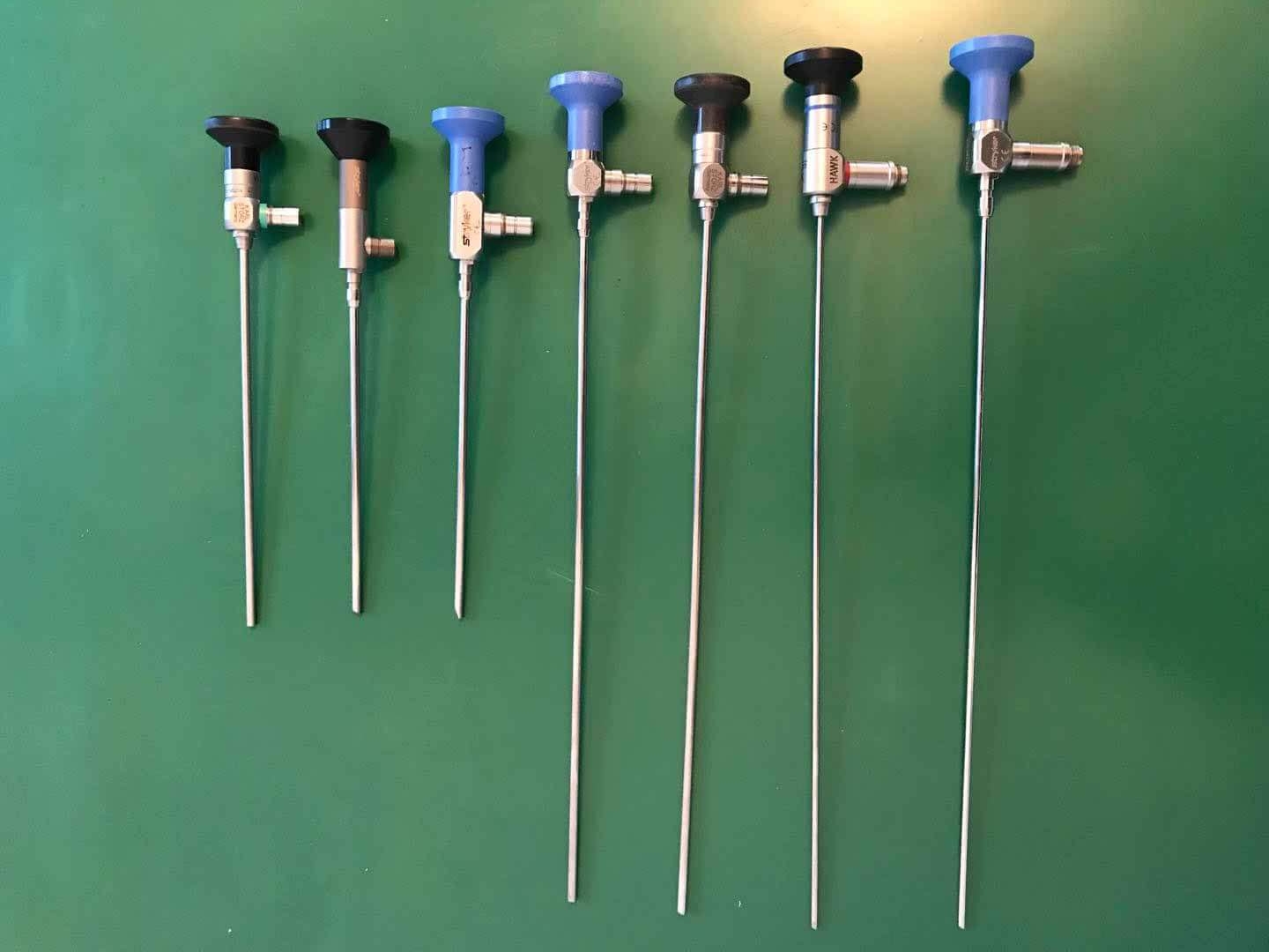
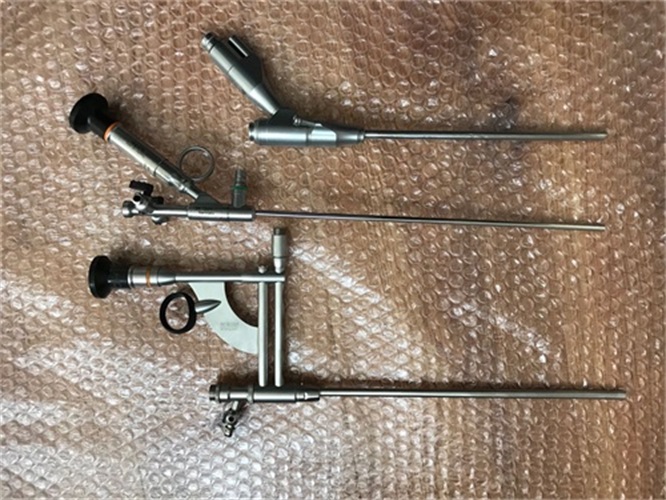
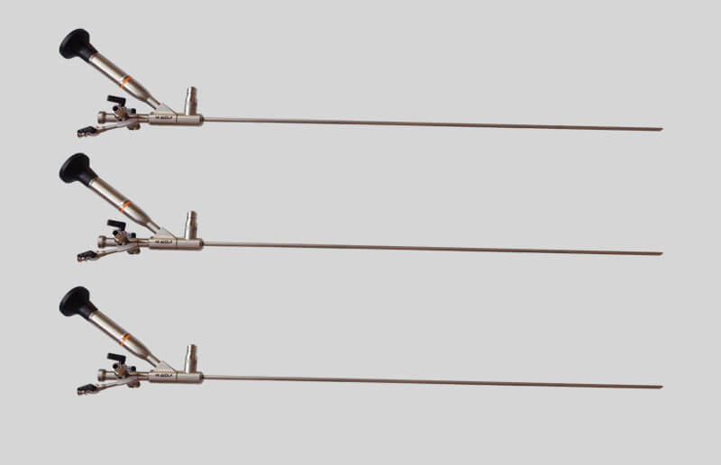
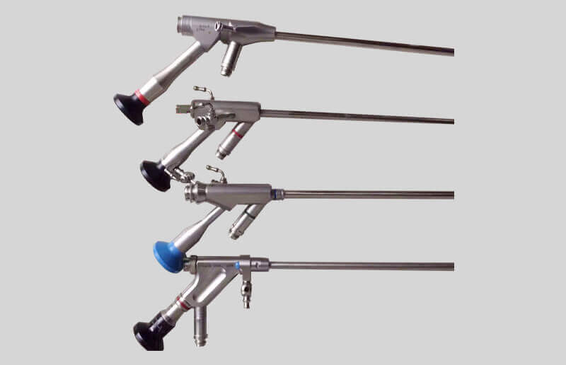
医用内窥镜主要分为以下两大类:硬管式内窥镜:不可弯曲,经过外科切口进入人体无菌组织、器官、无菌腔室,主要包括腹腔镜、胸腔镜、关节镜等,硬管式内窥镜可分为白光硬镜和荧光硬镜。软管式内窥镜:可以自由弯曲,通过人体自然腔道进入体内,镜体较长同时具有一定柔性,光电信号传输距离较远,镜体插入部直径较小且功能集成复杂,对设计工艺及制造技术的要求更高,具有较高的技术壁垒,主要包括胃镜、肠镜、支气管镜等。
Medical endoscopes are mainly divided into the following two categories: rigid tube endoscopes: non bendable, entering sterile tissues, organs, and chambers of the human body through surgical incisions, mainly including laparoscopy, thoracoscopy, arthroscopy, etc. Hard tube endoscopes can be divided into white light hard endoscopes and fluorescent hard endoscopes. Flexible tube endoscope: It can be freely bent and enter the body through the natural passage of the human body. The endoscope body is long and has a certain degree of flexibility, with a long distance for photoelectric signal transmission. The diameter of the insertion part of the endoscope body is small and the function integration is complex. It has higher requirements for design process and manufacturing technology, and has high technical barriers, mainly including gastroscopy, colonoscopy, bronchoscopy, etc.
医用内窥镜主要由三大系统组成,分别为窥镜系统、图像显示系统、照明系统。
Medical endoscopes mainly consist of three major systems, namely the endoscope system, image display system, and lighting system.

以目前较为常用的电子内窥镜为例,镜体伸入患者体内,镜体内部并列多个管道,包括照明光纤、传像光纤、传气通道、传水通道、器械通道等,内窥镜精密度极高,需要多个专业领域相互配合。
Taking the commonly used electronic endoscope as an example, the endoscope body is inserted into the patient's body, and multiple pipes are parallel inside the endoscope body, including lighting optical fibers, image transmission optical fibers, gas transmission channels, water transmission channels, instrument channels, etc. The precision of the endoscope is extremely high, requiring cooperation from multiple professional fields.
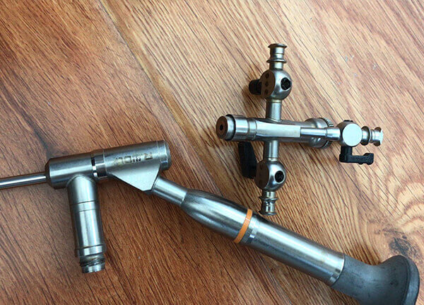
内窥镜系统:主要包括手柄和镜体。镜体主要由物镜、传像元件、目镜、照明元件及辅助元件等组成。图像显示系统:早期内窥镜或硬管式内窥镜采用直视,而现在的电子内窥镜通常由CCD/CMOS光电传感器、显示器、计算机和图像处理器组成。照明系统:主要是照明光源、传光束等。早的内窥镜设备采用热光源,如自然光、煤油灯、通电铂丝环、小型白炽灯等,易对人体造成灼伤,需要同时配置水冷装置,现在则普遍使用冷光源,主要是LED光源、氙灯、卤素灯。
Endoscopic system: mainly including handle and endoscope body. The mirror body mainly consists of an objective lens, image transmission components, eyepiece, lighting components, and auxiliary components. Image display system: Early endoscopes or rigid tube endoscopes used direct vision, while modern electronic endoscopes typically consist of CCD/CMOS photoelectric sensors, displays, computers, and image processors. Lighting system: mainly consisting of lighting sources, transmission beams, etc. Early endoscopic equipment used thermal light sources, such as natural light, kerosene lamps, electrified platinum wire rings, small incandescent lamps, etc., which could easily cause burns to the human body and required water cooling devices. Nowadays, cold light sources are commonly used, mainly LED light sources, xenon lamps, and halogen lamps.
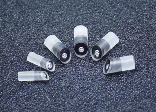
成像镜头:以往采用球面设计,使得像差和变形增大,结果会出现明显的影像不清,视界歪曲、视野狭小等不良现象,而现在的非球面设计是由球面和平面以外的曲面组成,通过改变镜片的曲率,使光线汇聚在固定的焦点,修正了影像,解决视界歪曲等问题,同时使镜片更轻、更薄、更平。图像传感器:图像传感器是感受光学图像信息并转换成可用输出信息的传感器,是组成数字摄像头的重要器件。可分为电荷耦合元件(CCD)和金属氧化物半导体元件(CMOS)。CCD高解析度、动态范围广、低失真度,但功耗大;CMOS体积小、能耗低、成本低、系统整合度高,但信噪比低、图像质量较差,国内外技术差距不大。
Imaging lens: In the past, spherical design was used to increase aberration and deformation, resulting in obvious image blurring, distorted field of view, narrow field of view, and other adverse phenomena. However, the current non spherical design is composed of spherical surfaces and surfaces outside the plane. By changing the curvature of the lens, light rays converge at a fixed focus, correcting the image, solving the problem of distorted field of view, and making the lens lighter, thinner, and flatter. Image sensor: Image sensor is a sensor that senses optical image information and converts it into usable output information. It is an important component of digital cameras. It can be divided into charge coupled devices (CCD) and metal oxide semiconductor devices (CMOS). CCD has high resolution, wide dynamic range, low distortion, but high power consumption; CMOS has small size, low energy consumption, low cost, and high system integration, but low signal-to-noise ratio and poor image quality, and there is not much difference in technology between domestic and international markets.
图像降噪:图像降噪是指减少数字图像中噪声的过程。在内窥镜摄像系统中,由于应用场景的特殊性,需针对微创手术的应用特点进行针对性的图像处理,以满足临床诊疗需求。在内窥镜运动时会产生模糊的图像,用降噪算法筛选出拍摄图像中与对象相关移动相对缓慢的像素呈现,从而保持图像的干净清晰,让医生可以做出准确的诊断;边缘增强技术是通过算法生成对比度强烈的血管视图,方便医生分析;使用假彩色成像、数字滤波等技术,可以把一些不明显的或是早期病变彰显出来。边缘增强:边缘增强的目的是提高图像的质量和可辨识度,使图像更有利于观察或进一步分析处理,帮助医生更全面查看组织中的非正常现象,也是内窥镜图像处理中非常重要的技术。
Image denoising: Image denoising refers to the process of reducing noise in digital images. In the endoscopic camera system, due to the special application scenarios, targeted image processing needs to be carried out according to the application characteristics of minimally invasive surgery to meet clinical diagnosis and treatment needs. During the movement of the endoscope, blurry images are generated. A denoising algorithm is used to filter out pixels in the captured image that move relatively slowly related to the object, in order to maintain the cleanliness and clarity of the image, allowing doctors to make accurate diagnoses; Edge enhancement technology generates blood vessel views with strong contrast through algorithms, making it easier for doctors to analyze; Using techniques such as false color imaging and digital filtering can highlight some inconspicuous or early lesions. Edge enhancement: The purpose of edge enhancement is to improve the quality and recognizability of images, making them more conducive to observation or further analysis and processing, helping doctors comprehensively examine abnormal phenomena in tissues, and is also a very important technology in endoscopic image processing.
例如根据颜色难以将细小的血管与周边组织区分开来,但可以采用边缘增强技术生成对比度较强烈的血管视图,以供医生分析之用。此外,边缘增强还常用于改进组织纹理图像以及黏膜表面图像的视图质量。
For example, it is difficult to distinguish small blood vessels from surrounding tissues based on color, but edge enhancement technology can be used to generate vascular views with stronger contrast for doctors to analyze. In addition, edge enhancement is often used to improve the view quality of tissue texture images and mucosal surface images.
本文由内窥镜设备维修友情奉献.更多有关的知识请点击:http://www.jiangrenyiliao.cn真诚的态度.为您提供为全面的服务.更多有关的知识我们将会陆续向大家奉献.敬请期待
This article is dedicated to the maintenance of endoscopic equipment. For more information, please click: http://www.jiangrenyiliao.cn Sincere attitude. We will provide you with comprehensive services. We will gradually contribute more relevant knowledge to everyone. Stay tuned
 新闻资讯
新闻资讯 联系我们
联系我们 公司:匠仁医疗设备有限公司
  新闻资讯
新闻资讯微信公众号

樊经理:13153199508 李经理:13873135765
公司地址:山东省济南市槐荫区美里东路3000号德迈国际信息产业园6号楼101-2室 湖南省长沙市雨花区劳动东路820号恒大绿洲14栋2409室
备案号:鲁ICP备2023027194号-1 鲁公网安备 37010402441281号