内窥镜是是具有观察目的的医学仪器,是一种光学装置,可以深入人体自然腔道或通过外科手术打开的孔道进行检查、诊断或治疗的医疗器械。
Endoscope is a kind of medical instrument with the purpose of observation. It is an optical device, which can go deep into the natural cavity of human body or through the opening of the hole for examination, diagnosis or treatment.
内窥镜主要由三大系统组成,分别为窥镜系统、图像显示系统、照明系统。以目前较为常用的电子内窥镜为例,窥镜系统包括手柄和镜体,镜体伸入患者体内,镜体内部并列多个管道,包括照明光纤、传像光纤(电子内窥镜中是CCD视频线)、传气通道、传水通道、器械通道等。
Endoscope is mainly composed of three systems, which are endoscope system, image display system and lighting system. Taking the electronic endoscope as an example, the endoscope system includes a handle and a mirror body, the mirror body extends into the patient's body, and there are several parallel pipes in the mirror body, including lighting optical fiber, image transmission optical fiber (CCD video cable in the electronic endoscope), gas transmission channel, water transmission channel, device channel, etc.
一、内窥镜发展情况及运用场景
1、 Development and application of endoscope
1、发展史
1. History of development
内窥镜的发展经历了硬管式内窥镜(1806-1932)、半曲式内窥镜(1932-1957)到纤维内窥镜(1957以后)、电子内窥镜(1983年以后)四个阶段。按其成像构造分类:可大体分为3大类:硬管式内窥镜、光学纤维(可分为软镜和硬镜)内窥镜和电子内窥镜(可分为软镜和硬镜)。
The development of endoscope has gone through four stages: hard tube endoscope (1806-1932), half curved endoscope (1932-1957), fiber endoscope (after 1957) and electronic endoscope (after 1983). According to its imaging structure, it can be roughly divided into three categories: hard tube endoscope, optical fiber (soft mirror and hard mirror) endoscope and electronic endoscope (soft mirror and hard mirror).
2、内窥镜运用场景
2. Endoscope application scene
二、内窥镜分类介绍
2、 Classification of endoscopes
1、硬管式内窥镜
1. Rigid tube endoscope
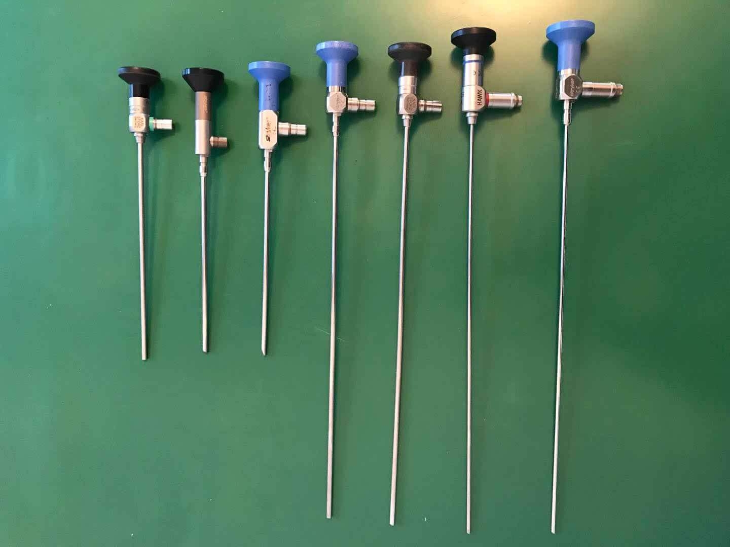
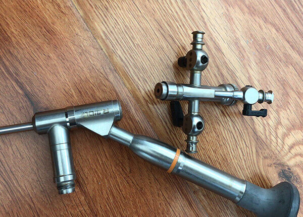
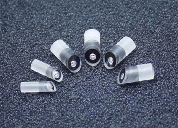
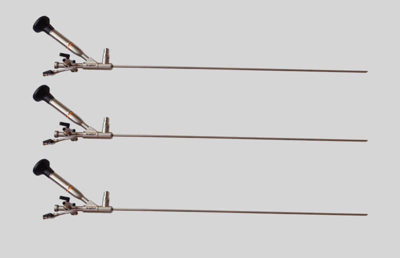
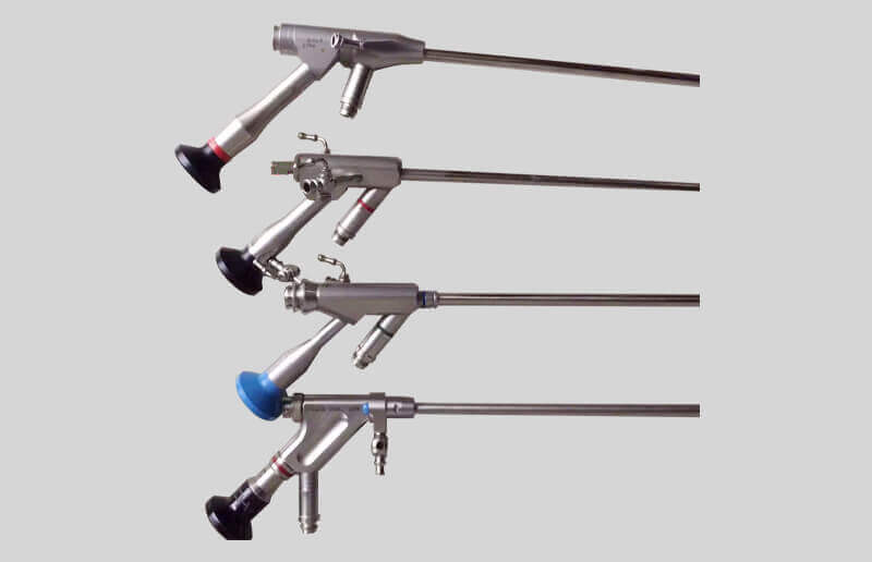
硬管内窥镜,主要用于人体表浅及浅层部位自然腔道和通过穿刺开口腔道的病灶诊断和(或)治疗,如膀胱镜、腹腔镜,在操作中不可弯曲。硬管内窥镜主要由光学成像系统和照明系统组成:光学部分外观看是一个细长的金属管子,而里面装着一个由许多透镜组成的完整的光学系统。光学成像系统由物镜系统、转像系统、目镜系统三大系统组成。
Hard tube endoscopy is mainly used for the diagnosis and / or treatment of the lesions in the natural lumen of the superficial and shallow parts of the human body and through the puncture of the open lumen, such as cystoscope and laparoscope, which cannot be bent during the operation. The hard tube endoscope is mainly composed of optical imaging system and lighting system: the optical part is a long and thin metal tube, and there is a complete optical system composed of many lenses inside. The optical imaging system is composed of objective system, image rotation system and eyepiece system.
2、纤维内窥镜
2. Fiberscope
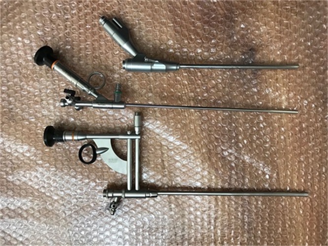
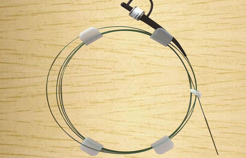
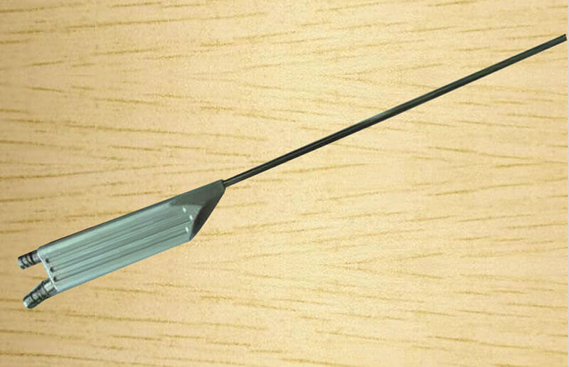
纤维内窥镜一般由目镜、手轮(软性或半硬性)、钳道口、导光束接口、导像束、导光束组成,有些产品还包括送水(气)孔、闭孔器等。纤维内窥镜由光学观察系统、照明传输系统和支架构件组成。光学观察系统由聚焦成像的物镜组、传输物镜组像的传/转像组和目视观察用的目镜或CCD转接镜构成;照明传输系统由混编排列的多束导光纤维构成;支架构件由支承并包裹前述系统并开有手术或冲洗孔道的医用金属或有机材料构成。
Fiber endoscope is generally composed of eyepiece, hand wheel (soft or semi-rigid), Tongdao port, light guide interface, image guide beam and light guide beam. Some products also include water (air) supply hole, hole closers, etc. The fiber endoscope consists of an optical observation system, a lighting transmission system and a bracket component. The optical observation system is composed of a focus imaging objective lens group, a transmission / conversion group for transmitting the objective lens group image and an eyepiece or CCD adapter for visual observation; the lighting transmission system is composed of a mixed arrangement of multi beam light guide fibers; the bracket component is composed of medical metal or organic materials supporting and wrapping the above system and opening an operation or washing channel.
纤维内窥镜按用途分为:上消化道内窥镜、下消化道内窥镜、呼吸道内窥镜。
Fiberscope can be divided into upper digestive tract endoscope, lower digestive tract endoscope and respiratory tract endoscope according to its application.
按光学视向角分为:前视型、斜视型、侧视型三种。
According to the optical viewing angle, it can be divided into three types: forward viewing type, squint viewing type and side viewing type.
按功能分为:具有手术功能(带手术和/或冲洗孔道)和不具有手术功能(检查用)两种。
According to the function, it can be divided into two types: with operation function (with operation and / or flushing hole) and without operation function (for examination).
3、电子内窥镜
3. Electronic endoscope
电子内窥镜主要由内镜、电视信息系统中心和电视监视器三个主要部分组成,另外还配备一些辅助装置,如录像机、照相机、吸引器以及用来输入各种信息的键盘和诊断治疗所用的各种处置器具等。
The electronic endoscope is mainly composed of three main parts: endoscope, TV information system center and TV monitor. In addition, it is also equipped with some auxiliary devices, such as video recorder, camera, attractor, keyboard for inputting various information and various disposal devices for diagnosis and treatment.
4、超声内镜
4. Endoscopic ultrasonography
将微型高频超声探头安置在内镜前端,在内镜直接观察腔内形态的同时,又可进行实时超声扫描,以获得管道壁各层次的组织学特征及周围邻近脏器的超声图像。
The micro high-frequency ultrasound probe is placed in the front of the endoscope. When the endoscope directly observes the shape of the cavity, it can also carry out real-time ultrasound scanning, so as to obtain the histological characteristics of the various layers of the pipe wall and the ultrasound images of the surrounding organs.
5、胶囊内镜
5. Capsule endoscopy
胶囊内镜最早由以色列科学家于 2000 年研发,胶囊内镜可以对全小肠进行拍照观察(胃镜和肠镜只能看到胃部和大肠,无法看到中间小肠这一段),弥补了胃镜和肠镜看不到的地方,大大提高了消化道疾病诊断检出率。
Capsule endoscopy was first developed by Israeli scientists in 2000. Capsule endoscopy can take photos of the whole small intestine (gastroscope and enteroscope can only see the stomach and large intestine, but can't see the middle small intestine). It makes up for the places that gastroscope and enteroscope can't see, and greatly improves the detection rate of digestive tract disease diagnosis.
胶囊胃镜需实现对胶囊的主动控制,目前主要有两种方式实现主动控制:内部驱动和外部驱动。内部驱动模式暂时缺乏可行性,外部驱动的主流方式是依靠体外磁场控制。目前主要有三大类磁控方法:手柄式、磁共振(MRI)线圈式和机器臂式磁控,目前仅机器臂式磁控获批用于临床胃部检查。根据磁控的类型,主要分为电磁体、永磁体和磁共振。
Capsule gastroscope needs to realize the active control of capsule. At present, there are two main ways to achieve the active control: internal drive and external drive. The internal driving mode is temporarily lack of feasibility, and the main way of external driving is to rely on external magnetic field control. At present, there are three main types of magnetic control methods: handle type, magnetic resonance (MRI) coil type and machine arm type. At present, only machine arm type magnetic control has been approved for clinical gastric examination. According to the type of magnetic control, it is mainly divided into electromagnet, permanent magnet and magnetic resonance.
三、内窥镜市场与技术
3、 Endoscope market and technology
1、内窥镜市场
1. Endoscope Market
2015 年全球内窥镜市场规模已经达到 302 亿美元,2011 年-2015 年复合增长率为 7.2%。2017 年国内市场规模约为 246 亿人民币,其中软性内窥镜的市场规模大约 33-35 亿人民币。从全球看软镜技术基本被日本主要几家企业垄断。奥林巴斯、宾得、富士合计占据 90%以上的市场份额,其中奥林巴斯市场份额超过 65%。我国硬镜市场一梯队为 Karl Storz 和奥林巴斯,二梯队为史塞克和狼牌,合计占据 85%以上的市场份额。国产硬镜的主要厂家包括沈阳沈大、好克和天松等,国产企业合计市场份额 5%左右。
In 2015, the global endoscope market has reached 30.2 billion US dollars, with a compound growth rate of 7.2% from 2011 to 2015. In 2017, the domestic market size was about 24.6 billion yuan, including 3.3-3.5 billion yuan for soft endoscopes. From a global perspective, soft mirror technology is basically monopolized by several major Japanese enterprises. Olympus, Pentax and Fuji together account for more than 90% of the market share, of which Olympus accounts for more than 65%. The first echelon of China's hard mirror market is Karl Storz and Olympus, and the second echelon is Stryker and wolf, accounting for more than 85% of the total market share. The main manufacturers of domestic hard mirror include Shenyang Shenda, Haoke and Tiansong. The total market share of domestic enterprises is about 5%.
2、内窥镜技术方向
2. Technical direction of endoscope
目前电子内窥镜的发展趋势为两方面:1、高清分辨率,目前已经达到百万像素;2、探头微型化。探头的大小直接影响创伤的严重程度,微型探头化可以减轻患者痛苦和不适度。
At present, the development trend of electronic endoscope is two aspects: 1. High resolution, which has reached one million pixels; 2. Probe miniaturization. The size of the probe has a direct impact on the severity of the trauma. Miniaturization of the probe can reduce the pain and discomfort of patients.
The above is a detailed introduction to the development history and classification of endoscopes. If you want to know more, please consult the endoscope equipment maintenance website http://www.jiangrenyiliao.cn 。

 新闻资讯
新闻资讯 联系我们
联系我们 公司:匠仁医疗设备有限公司
  新闻资讯
新闻资讯