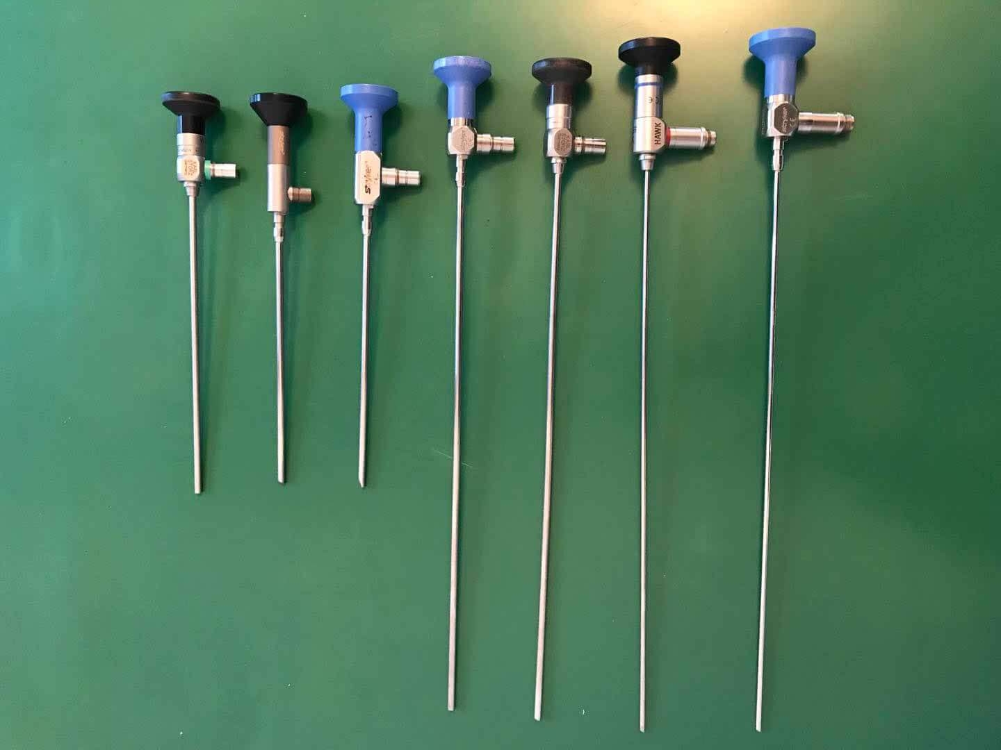
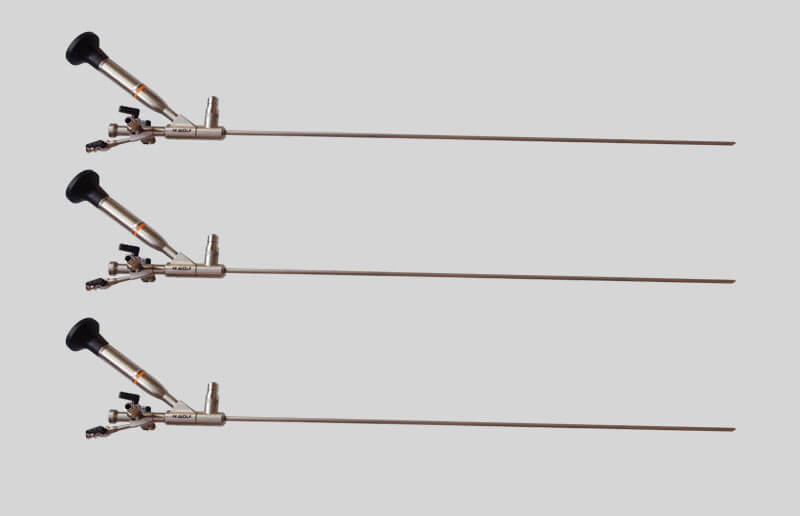
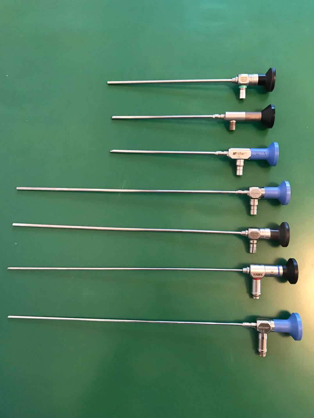
1、纤维内窥镜
1. Fiberscope
纤维内窥镜体系由内窥镜镜体和冷光源两部分组成,镜体内有两条光导纤维束: 一条叫光束,它是用来将冷光源发生的光线传导到被观测的物体外表,将被观测物外表照亮;另一条叫像束,它是把数万根直径在1微米以下的光导纤维按一行一行顺序摆放成一束,一端对准目镜,另一端经过物镜片对准被观测物外表,医师经过目镜能够非常直观地看到脏器外表的状况,便于及时准确地确诊病情。例如,凭借内窥镜医师能够调查胃内的溃疡或肿瘤,据此制定出最佳的治疗计划。
The fiber endoscope system consists of two parts: the endoscope body and the cold light source. There are two optical fiber bundles in the endoscope body: one is called the light beam, which is used to conduct the light generated by the cold light source to the surface of the observed object and illuminate the surface of the observed object; The other is called image beam. It is a bundle of tens of thousands of optical fibers with a diameter of less than 1 μ m placed in line by line. One end is aimed at the eyepiece, and the other end is aimed at the appearance of the observed object through the object lens. The doctor can see the appearance of the organs directly through the eyepiece, which is convenient for timely and accurate diagnosis of the disease. For example, endoscopists can investigate gastric ulcers or tumors to develop the best treatment plan.
传导图画的纤维束构成了纤维内镜的中心部分,它由数万根极细的玻璃纤维组成,依据光学的全反射原理,一切玻璃纤维外面有必要再被覆一层折射率较低的膜,以确保一切内芯纤维传导的光线都能发生全反射。单根纤维的传递只能发生一个光点,要想看到图画,就有必要把很多的纤维集成束,要想把图画传递到另一端也成相同的图画,就有必要使每一根纤维在其两端所摆放的位置相同,称为导像束。纤维内窥镜一般有两个玻璃纤维管,光经过其中之一进入体内,医师经过另一个管或经过一个摄像机来进行调查。
The fiber bundle conducting the picture forms the central part of the fiber endoscope, which is composed of tens of thousands of extremely fine glass fibers. According to the principle of total reflection of optics, it is necessary to cover a layer of film with low refractive index on the outside of all glass fibers to ensure that all the light conducted by the inner core fibers can have total reflection. The transmission of a single fiber can only take place at one light spot. To see a picture, it is necessary to integrate many fibers into a bundle. To transfer a picture to the other end into the same picture, it is necessary to place each fiber in the same position at both ends of the bundle, which is called the image guide bundle. Fiberoptic endoscopes usually have two fiberglass tubes. Light enters the body through one of them. Doctors conduct investigations through another tube or through a camera.
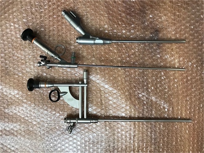
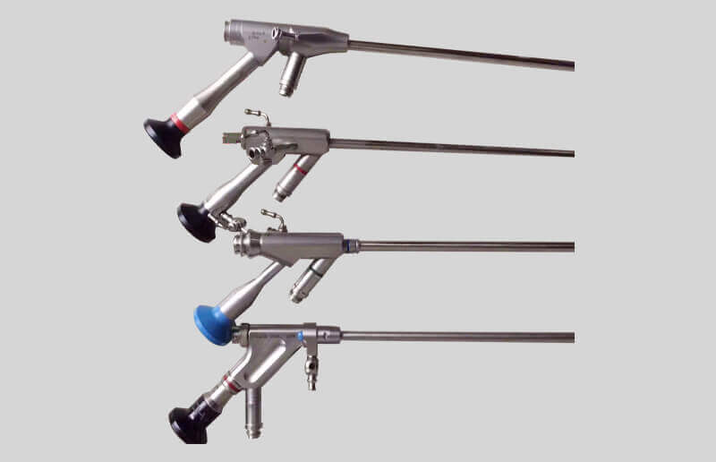
2、电子内窥镜
2. Electronic endoscope
随着电子学和数字视频技术的发展,与80年代出现了电子内窥镜,这样便不再以光纤传像,而代之以光敏集成电路摄像体系,简称CCD。微型图画传感器的CCD 器件是电荷耦合器件,是在硅基片上制成的大规模面阵集成电路芯片,是一种全固态成像器件。CCD芯片凭借必要的光学体系(内窥镜先端物镜)和专用的外围驱动与信号处理电路,能够将景物图画经过CCD面阵进行逐点、逐行、逐帧顺次转化 、存储 、传输,在其输出端发生一个景物图画相关的时序视频信号经电缆传输至外部电路转化处理体系经取样、A/D 转化、数字信号处理、D/A转化、电视信号编码,最终在监视器上还原成可供调查的景物图画和相关文字信息。首要所能显现的不但影像质量好,光亮度强,并且图画大,能够检查出更细微的病变,并且电子内窥镜的外径更细,图画更加明晰和直观,操作便利。有些内窥镜甚至还有微型集成电路传感器,将所调查到的信息反馈给计算机。它不但能取得安排器官形态学的确诊信息,并且也能对安排器官各种生理机能进行测定。With the development of electronics and digital video technology, electronic endoscope appeared in 1980s, so it is no longer transmitted by optical fiber, but instead by light-sensitive integrated circuit camera system, referred to as CCD. The CCD device of the miniature picture sensor is a charge coupled device, a large-scale array integrated circuit chip made on a silicon substrate, and an all solid-state imaging device. With the necessary optical system (endoscope first objective) and special peripheral drive and signal processing circuit, CCD chip can transform, store and transmit the scene pictures point by point, line by line, frame by frame through CCD array, and generate a sequential video signal related to the scene pictures at its output end, which is transmitted to the external circuit conversion processing system through cable, and then through sampling, a / d Conversion, digital signal processing, D / a conversion, TV signal coding, and finally restore the scene pictures and related text information available for investigation on the monitor. What can be shown above all is not only good image quality, strong brightness, but also large picture, which can detect more subtle lesions, and the outer diameter of the electronic endoscope is smaller, the picture is more clear and intuitive, and the operation is convenient. Some endoscopes even have micro integrated circuit sensors that feed the information back to the computer. It can not only obtain the diagnosis information of the arrangement organ morphology, but also measure the physiological functions of the arrangement organ.
电子内窥镜与纤维内窥镜的本质区别
The essential difference between electronic endoscope and fiber endoscope
电子内窥镜构造与纤维内镜构造基本相同,简略可理解为用 CCD 代替了导像束,很多功用是纤维内窥镜不能企及的。电子内窥镜与纤维内窥镜比较最大的不同之处是用被称为微型图画传感器的CCD器件取代了光导纤维传象束。
The structure of the electronic endoscope is basically the same as that of the fiber endoscope. It can be simply understood that CCD is used to replace the image guide beam, and many functions cannot be achieved by the fiber endoscope. The biggest difference between the electronic endoscope and the fiber endoscope is that the CCD device called the miniature picture sensor is used instead of the optical fiber image beam.
另:
The other:

 新闻资讯
新闻资讯 联系我们
联系我们 公司:匠仁医疗设备有限公司
  新闻资讯
新闻资讯